If knowledge about genetics of human diseases is available, it can be used in a variety of ways to avoid or reduce the incidence of some of these diseases. This can be achieved in a variety of ways and we will describe five of them, namely (i) genetic counselling, (ii) antenatal diagnosis, (iii) gene therapy, (iv) making choice of baby's sex, and (v) DNA fingerprinting in forensic science.
Genetic counsellingGenetic counselling for couples who believe that there may be a risk of producing a defective child, has now become routine aspect of medical practice, particularly in the developed countries. These parents may either voluntarily abstain from having any child or may undergo selective abortions on suspicion or after ascertaining it through antenatal diagnosis.
A genetic counsellor should first be able to identify the disease and therefore should be first a clinician and then a geneticist. The simplest cases asking for genetic counselling will be those having a family history of disease and the parents may like to know the chances of having a child free of that disease. A couple may have one defective child and would like to know the chances of having a normal child on the next pregnancy. In such a case, if the defect is known to be single gene recessive and both parents are normal, the chance is three in four of having a normal child, although even a normal child will have a two third chance of being a carrier. The parents may like to give birth to such a child who may be a carrier, because the chance of his or her spouse also being a carrier will be remote. However, in such cases, even the possibility of having the defective grand child can be worked out, if the frequency of heterozygotes in the population available for marriage of the child is known. For instance, in case of fibrocystic disease (cystic fibrosis) of the pancreas, the frequency in general population (perhaps in UK) is 1/22. The chance of the normal child to be a carrier being 2/3 and the chance of spouse to be a carrier being 1/22, the chance of the grand child to be defective would be 2/3x 1/22 x 1/4 = 1/l32 (since 1/4 is the chance that a child born to both heterozygote parents will be defective). A risk of one in 132 may or may not be worth taking depending upon temperament and circumstances. More detailed calculation can be done in these simple cases and also in cases of polygenic nature and variable penetrance. Readers are advised to consult a book on human genetics for further details.
In some cases, detection of heterozygote, may also be useful and possible. Following are the situations where it is possible, (i) When a heterozygote, though phenotypically normal, produces a particular enzyme activity intermediate to those found in two homozygotes, its presence can be detected using electrophoresis. In such cases, if a biochemistry laboratory is available, deficiency like HGPRT (Lesch-Nyhan syndrome) in heterozygous condition can be detected from a blood sample or some skin cells, (ii) When the mutant produced an altered form of gene product, the heterozygote may produce two different forms of protein that can be separated by electrophoresis to enable the identification of a heterozygote. (iii) If the defect is associated with a chromosome structure, then with the availability of a cytogenetics laboratory, such an abnormality in heterozygous condition can be identified.
Amniocentesis and antenatal diagnosisWhen a pregnant woman is known to have a chance of bearing a child with a genetic defect, it may be desirable to diagnose the condition in the fetus. This can be done by taking some cells from the fetus by drawing a few milliliters of amniotic fluid with the help of a hypodermic needle; The technique is called amniocentesis and is usually performed at 15th week of pregnancy, to allow enough time for safe abortion if recommended. The amniotic fluid has free cells of fetal origin, which can be cultured and tested in various ways e.g. karyotype, enzyme production and restriction site pattern analysis of its DNA. At least 35 diseases which can be identified by this technique are known. If disease is detected through such an antenatal diagnosis, abortion of fetus can then be recommended. However, if abortion is not acceptable to parents, there is no point in carrying out antenatal diagnosis.
Technique of amniocentesis, used to test for hereditary or developmental defects in the fetus
It is possible to identify the disease now within 2 months of pregnancy, unlike an 18 week period required earlier. The number of disease specific DNA probes is also increasing at a fast rate, so that antenatal diagnosis by DNA analysis or linkage should be possible for all single gene defects. In recent years, the incidence of the disease thailassaemia in Cypriot community in Britain has fallen from 30 to 2 per year, due to the use of antenatal diagnosis. In U.S.A. on the other hand, there is a campaign against abortion and, therefore, also against antenatal diagnosis. In such cases there will be births of defective children and these may become patients for gene therapy discussed in the following section.
Gene therapyIf a child is diagnosed to carry a defective gene leading to disability, one may like to get this gene replaced by a normal functional gene. This is gene therapy in theory. One would like to ask that if there is a need and demand for gene therapy, can it be done ? The answer to this question has changed from 'no' to 'yes' in recent years. The possibilities are being explored and treatment by transfusion of cells with functional gene, if not by replacement of defective gene, are being suggested and tried. Gene therapy can be used at two different levels : (i) patient therapy, in which cells with healthy gene may be introduced in the affected tissue, so that the healthy gene overcomes the defect without affecting the inheritance of the patient and (ii) embryo therapy, in which the genetic constitution of embryo at the post-zygotic level is altered, so that the inheritance will be altered. It is believed that in future, gene therapy of both types will possible.
Patient therapy. Patient therapy will involve the following steps : (i) the defective gene should be identified, (ii) normal healthy gene should either be isolated or synthesized, (iii) isolation of cells of the tissue, where the normal healthy gene will need to function, (iv) the normal gene should be placed into a cell, where it can function. The gene will have to be placed into the correct site on the host chromosome, so that the gene may function, or even one may have to delete the defective gene. There are three main problems in this connection. First, the introduced gene may not function, second, that when corrected cells are reintroduced, these may be outnumbered by the non-cured resident cells, and third, there are only few diseases affecting only a single tissue.
Utilizing the above approach, first clinical gene transfer (approved in U.S.A.) was achieved in 1989. It was a marker gene (neomycin resistance = NeoR) introduced into tumor-infiltrating lymphocytes (TIL). These NeoR/TILs were transferred into patients with advanced cancer, to ensure that the approved protocol really works. The first approved gene therapy protocol for correction of adenosine deaminase (ADA) deficiency, however, began in 1990 and by the end of 1992, two dozen active clinical protocols on three continents became available for trials. However, the technology is still very expensive and specialized to be used extensively. Other less expensive techniques (involving delivery of gene through vectors) are being developed.
Embryo therapy. This will involve the following steps, which have been tried in case of mouse or rabbit only, (i) in-vitro fertilization of the egg. (ii) insertion of normal gene into embryo at post zygotic level, either with viruses or directly by microinjection, (iii) integration of inserted gene in host DNA, where it may or may not function. The inserted genes have been found to be inactive generally, but in few animals, genes have been switched on in a tissue specific way but their activity is at a very low level. However, it is not yet possible that the therapeutic newly inserted genes function under normal control in the animal, in time, space or quantity.
Making a choice of baby's sexMarried couples have always been interested in knowing the sex, and sometimes in making a choice about the sex, of their babies. The chromosome techniques have made it possible to know the sex of developing fetus by drawing amniotic fluid and preparing karyotypes from cells derived from the fetus and floating in this fluid. There are clinics available now which can advise pregnant ladies about the sex of the developing fetus, so that the ladies can decide early about the abortion of fetus, if it belongs to the unwanted sex.
Recently techniques have also been developed, which will not require preferential abortion but will allow preferential fertilization by male (carrying Y chromosome) or female (carrying X chromosome) determining sperms. There are techniques available now, which allow separation of sperms carrying Y chromosomes, from the ejaculate of a man (through Ericson's method developed by R. Ericson of U.S.A.) to be used for insemination of an ovulating woman. This technique (using quinacrine stain) has been used in more than 50 sperm centres in the world including some in India (one in Bombay at Khar Road), with 80% success. Ericson has actually established a company named Gametrics Ltd. in California, U.S.A. which specializes in separating sperms with Y chromosome and hundreds of male children have been produced with its help.
Techniques have also been developed to separate sperms carrying X chromosome for artificial insemination leading to the birth of female children. This technique involves the use of sephdex gel column, in which sperms with Y, being lighter are trapped in gel and those with X being heavier reach the bottom of the column, and can be used for insemination.
The techniques permitting choice of sex of the baby have been condemned by many sociologists, who fear that this may disturb the sex ratio leading to a variety of problems. But some doctors argue that this will help couples in planning their families, since there are also couples who may like to have a female child. This may also allow selection against sex linked abnormalities in the children.
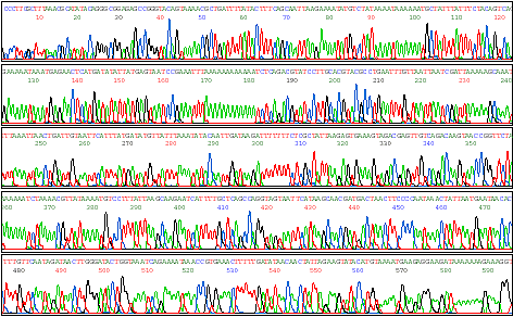
DNA fingerprinting in forensic scienceThe technique of DNA fingerprinting was developed for the first time (1985, 86) by Alec Jeffreys and his colleagues at Leicester University in U.K. In this field, establishment of the identity of a person with the help of blood stains, semen (sperms) stains or hair roots will be possible with almost absolute certainty. In this technique, DNA will be isolated from blood stains, semen stains or hair roots and will be subjected to Southern blotting and DNA hybridization with the help of specific DNA probes. This will reveal polymorphism in DNA, which has a very stable inheritance. For this purpose, DNA from blood, semen or urine stains may also be amplified using PCR technique (consult Genetic Engineering and Biotechnology 1. Recombinant DNA and PCR (Cloning and Amplification of DNA)).
It is speculated that the above technique will allow the identification of rapists in rape cases, and of mother and or father in case of doubtful parentage (Fig. 24.9). This technique will allow identification even when the stains on victim's clothes etc. are several years old and with much more certainty than has hitherto been possible through techniques of blood groups etc., since the number of blood groups available becomes a limitation. The technique of DNA fingerprinting reveals such a great polymorphism that the possibility of two persons having same pattern of DNA fingerprints is very remote.
DNA fingerprints, showing hypervariable sequences inherited by identical twins from their mother (M) and father (M). Arrows indicate bands common with father, but absent in mother (after A.J. Jeffreys, 1985).
In India, DNA fingerprinting tests are carried out at the Centre for Cell and Molecular Biology (CCMB), Hyderabad. For this purpose, a test with the Bkm probe (banded krait minor satellite DNA) earlier used for identification of sex chromosomes (consult Sex Determination, Sex Differentiation, Dosage Compensation and Genetic Imprinting), has been found to cost one-tenth of the cost of tests used in Europe and U.S.A. Paternity dispute cases are much more common in India-and most of them are referred to CCMB for DNA evidence. The first such test on DNA fingerprinting was used in June, 1989 to settle a drawn-out paternity case in Madras.